Subject: Protein Stucture and Function Wed Sep 22, 2010 12:26 pm
Proteins are extemely important molecules in living organisms which have a wide range of important functions. Your forum task is too investigate the structure and function of one protein.
1) Re-read the textbook section on proteins for back ground information
2) Watch the video below for further examples of possible structures
3) Research on the internet and find one example of a protein which is important in living organisms. You must attach a picture and also write a summary of at least 150 words about the structure and function of the protein.
IMPORTANT: You must not choose any protein from the textbook. You must not choose any protein already selected by a classmate and included in the forum.
DUE DATE: You must post your forum by the start of your second class during Week 29 (11/10/10)
Fran Martinez
Posts : 2 Join date : 2010-08-08
Subject: Re: Protein Stucture and Function Mon Oct 11, 2010 8:51 pm
Structure: Collagen is a fibrous protein; that is, it is composed of many fibers. Each fiber consists of three microscopic ropes of protein wrapped around each other. The fibers in collagen are arranged parallel to each other, and are often grouped together in bundles. The bundling of collagen fibers gives the fibers greater strength than if they occurred individually. Collagen fibers are extremely tough and can resist a pulling force, but because they are not taut, they allow some flexibility.
Functions: Collagen is a primary component of the connective tissue located in the dermis, the tough inner layer of the skin. This kind of connective tissue is also found in mucous membranes, nerves, blood vessels, and organs. Collagen in these structures imparts strength, support, and a certain amount of elasticity Collagen adds strength to tendons and ligaments, and it imparts some stretch to these structures by allowing for some flexibility. However, collagen is not extremely elastic. If tendons and ligaments are stretched too far, these structures will tear, which may lead to problems in movement and bone position. Many athletes tear tendons and ligaments. When tearing occurs, the joint or bone in which the structures occur must be immobilized to allow for proper healing. Collagen is also a component of a kind of connective tissue that surrounds organs. This connective tissue encases and protects delicate organs like the kidneys and spleen.
http://www.3dchem.com/molecules.asp?ID=195
Andrea G.
Posts : 2 Join date : 2010-06-30
Subject: Andrea G. Tue Oct 12, 2010 10:15 pm
Protein:Human serum albumin Human serum albumin is the most abundant protein in human blood plasma. It is produced in the liver. Albumin comprises about half of the blood serum protein. It is soluble and monomeric. Functions: • Maintains oncotic pressure • Transports thyroid hormones • Transports other hormones, particularly ones that are fat soluble • Transports fatty acids to the liver • Transports unconjugated bilirubin • Transports many drugs; serum albumin levels can affect the half-life of drugs • Competitively binds calcium ions (Ca2+) • Buffers pH • Serum albumin, as a negative acute-phase protein, is down-regulated in inflammatory states. As such, it is not a valid marker of nutritional status; rather, it is a marker in inflammatory states • Prevents photodegradation of folic acid Structure Human serum albumin is a single peptide chain of 585 amino acids, held in three homologous domains by 17 disulfide bonds. Within each domain are two long loops plus one shorter loop. The S-S bonds provide stability while the intervening peptide strands allow for flexibility. The configuration includes 67% alpha helix and 10% beta turn.
soli gutierrez
Posts : 2 Join date : 2010-08-06
Subject: Re: Protein Stucture and Function Tue Oct 12, 2010 10:36 pm
structure keratin is made of coiled polypeptides like cysteine disulphide which coiled together by disulphide bridges which forms a helix like structure very strong. The atoms of sulphur join together across this helix making a fibrous matrix which is difficult to brek or dissolve. The keratin may be very tough or soft and that is determined by the amount of cysteine disulphide the keratin contains. If the keratin has a lot of it then it is going to be hard like hooves (due to the sulphur in it) and if it has little amount tha keratin will be soft (like hair or skin).
funtion keratin is made of many keratynocytes wich are living cells that form skin, hair, nails and other parts of the body that contain keratin. It plays a very important role as what it forms is really important, like skin. Skin helps prevent diseases or infections due to bacteria in enviroment, and it protects muscles and organs from external damage. Hair is made of keratin also and it is very important for the mantainance of body temperature (insulation), in this way it helps homeostasis.
http://itech.dickinson.edu/chemistry/?cat=69
dtabak
Posts : 2 Join date : 2010-08-08
Subject: Titin protein Wed Oct 13, 2010 8:59 pm
Protein: Titin
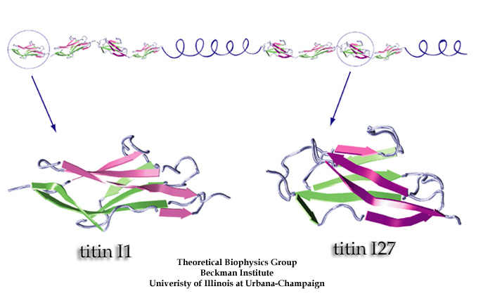
Titin is a protein in the human body, located especially in the cardiac and skeletal muscles. Its’ function is to allow passive elasticity in the muscles by acting as a molecular spring. Titin contracts and relaxes allowing muscle to expand and come back to normal position without becoming loose, as an elastic spring would. It is the largest single polypeptide ever found, it is 34350 amino acids long. Titin protein is made of two types of domains arranged in a line. The first domain is fibronectin type 3 domains and the second one is immunoglobulin domain. This linear arrangement is further organized in two regions: 1. N-terminal: elastic part of the molecule, mainly made of immunoglobulin domains 2. C-terminal: it’s made of both domains. It poses kianse activity. This protein contains Beta It’s a tertiary structure because of the bending and twisting of its domains.
Subject: Creatine Phosphokinase Thu Oct 14, 2010 12:19 am
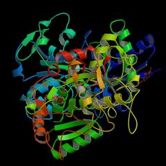
Creatine phosphokinase (CPK) is an enzyme found in the heart, brain and skeletal muscle. This protein intervenes in the biochimical reaction needed to creat energy in order to create nerve impulses. It catalyses phosphocreatin so it donates its phosphate to the ADP molecule, turning it into an ATP making it a new reserve of chemical energy Ready to be turned into mechanical energy needed for the process of contracting a muscle. There are three types of CPK that exist: 1: CPK-1 or CPK-BB present in the brain tissue nad lungs 2: CPK-2 or CPK-MB present in the heart 3: CPK-3 or CPK-MM present in the sekeletal muscles Laura Andrighetti
Emily Lynch
Posts : 2 Join date : 2010-06-30
Subject: Re: Protein Stucture and Function Thu Oct 14, 2010 3:01 pm
Myosin is not one protein, but a group of proteins found in eukaryotic tissues. They move along the actin filaments, hydrolizing ATP and are responsible for actin based molity. They share the basic properties of actin binding and force transduction. There are more than 20 different types of myosin that differ in their motor and tail domains (structure). Due to different structure, myosin fulfill different jobs, though all of them related to their main function. For example myosin II can generate force in skeletal muscle by realeasing phosphates from the myosin molecules. Myosin are made up of three "domains": head, neck, and tail domain. Myosin I: made up of one heavy chain, with one globular motor domain and a relatively short tail Myosin II: includes two heavy chains and towl light chains. The heavy chains have a globular motor domain and two alpha helix tail domains. The light chains are called either a regulatory chain or an essential chain.
vanessa hites
Posts : 1 Join date : 2010-10-14
Subject: vanessa Thu Oct 14, 2010 8:26 pm
Myosin is one of the two major protein constituents responsible for contraction of muscle. In muscle cells myosin is arranged in long filaments called thick filaments that lie parallel to the microfilaments of actin. In muscle contraction, filaments of actin alternately chemically link and unlink with those of myosin in a creeping or sliding action. The energy for this reaction is supplied by adenosine triphosphate. Myosin and actin also function in the motility of diverse non-muscle cells. In slime molds, for example, although present in much smaller quantities and forming shorter filaments, the interaction of the two proteins is employed to change cell shape and permit some movements.
cant attach the photo sorrry, there is the link
mikel villalabeitita
Posts : 2 Join date : 2010-08-12
Subject: Re: Protein Stucture and Function Thu Oct 14, 2010 10:28 pm
Cadherins (named for "calcium-dependent adhesion") are a class of type-1 transmembrane proteins. They play important roles in cell adhesion, ensuring that cells within tissues are bound together. They are dependent on calcium (Ca2+) ions to function, hence their name.
The cadherin superfamily includes cadherins, protocadherins, desmogleins, and desmocollins, and more.In structure, they share cadherin repeats, which are the extracelular Ca2+-binding domains. There are multiple classes of cadherin molecule, each designated with a prefix (in general, noting the type of tissue with which it is associated). It has been observed that cells containing a specific cadherin subtype tend to cluster together to the exclusion of other types, both in cell culture and during development.[citation needed] For example, cells containing N-cadherin tend to cluster with other N-cadherin expressing cells. However, it has been noted that the mixing speed in the cell culture experiments can have an effect on the extent of homotypic specificity. In addition, several groups have observed heterotypic binding affinity (i.e., binding of different types of cadherin together) in various assays. One current model proposes that cells distinguish cadherin subtypes based on kinetic specificity rather than thermodynamic specificity, as different types of cadherin homotypic bonds have different lifetimes.
Teresita Eyzaguirre S
Posts : 2 Join date : 2010-08-07
Subject: Re: Protein Stucture and Function Thu Oct 14, 2010 11:07 pm
TRANSFERRIN: FUNCTION Transferrin is a blood plasma protein for iron delivery that, in humans, is encoded by the TF gene. Transferrin is a glycoprotein that binds iron very tightly but reversibly. Although iron bound to transferrin is less than 0.1% of the total body iron, it is the most important iron pool, with the highest rate of turnover. Transferrin has a molecular weight of around 80 kDa. Transferrin is also associated with the innate immune system. Transferrin is found in the mucosa and binds iron, thus creating an environment low in free iron, where few bacteria are able to survive. The levels of transferrin decreases in inflammation, seeming contradictory to its function.
STRUCTURE In humans, transferrin consists of a polypeptide chain containing 679 amino acids. It is a complex composed of alpha helices and beta sheets to form two domains (the first situated in the N-terminus and the second in the C-terminus). The N- and C- terminal sequences are represented by globular lobes and between the two lobes is an iron-binding site. The amino acids which bind the iron ion to the transferrin are identical for both lobes; two tyrosines, one histidine, and one aspartic acid. In order for the iron ion to bind an anion is required, preferably carbonate.
A deficiency is associated with atransferrinemia. A high transferrin level may indicate iron deficiency anemia. Levels of serum iron and total iron binding capacity (TIBC) are used in conjunction with transferrin to specify any abnormality.
[img][/img]
fdofantini
Posts : 2 Join date : 2010-08-19
Subject: Cadherins Sat Oct 16, 2010 6:40 pm
Cadherins: This type of proteing is basically dependent of calcium. As well as desmogleins, desmocollins, and protocadherins, this protein is part of the cadherin superfamily. Cadherin plays the role of making sure that the cells are bound together with the tissues. You can find a high variety of classes of cadherin molecules and all of these can be defined as similar as they all share similar characteristics. In time, it has been discovered that cells that share an specific cadherin subtype end by clusting together and excluding types that are diferent.(during the developnment and in the cell). Lately it is said that cadherin and the members of the cadherin superfamily are related to the kinetic type of specificity and not in the thermo specificy and have a diferent lifetime than other types of cadherin bonds. Structure: - In terms of structure cadherin and the other proteins of the cadherin superfamily, share cadherin reapeats. This mean that all of these share the extracellular ("Ca2+").
MISS NO PUDE PONER LA FOTO (NO SOY MUY BUENO CON ESTO DE LA TECHNOLOGIA) le dejo el link a la foto. - http://www.sciencecodex.com/structure_of_innerear_protein_is_key_to_both_hearing_and_inherited_deafness ( hay 2 )
franco bozzalla
Posts : 2 Join date : 2010-08-07
Subject: Re: Protein Stucture and Function Sat Oct 16, 2010 7:00 pm
keratin, any one of a class of fibrous protein molecules that serve as structural units for various living tissues. The keratins are the major protein components of hair, wool, nails, horn, hoofs, and the quills of feathers. These proteins generally contain large quantities of the sulfur-containing amino acids, particulary cysteine. The helical keratin molecules twist around each other to form elongated strands called intermediate filaments. The formation of a covalent chemical bond called a disulfide bridge between the sulfer atoms on two cysteins on separate polypeptide chains of keratin allows for the cross-linkage of these chains and results in a fairly rigid aggregate. This phenomenon is seen to be consistent with the physiological role of the keratins, which provide a tough, fibrous matrix for the tissues in which they are found. Human hair is approximately 14% cystine (cysteins cross-linked by disulfide bridges).
fernandamontes
Posts : 2 Join date : 2010-08-18
Subject: ACTIN Sun Oct 17, 2010 9:13 pm
Actin is a protein that is present in all eukaryotic cells. It is considered as a “highly-conserved” protein, this means it is a conserved gene which has ‘lived’ for a very long period. It is as well the subunit of the microfilament found in cytoskeleton (cellular skeleton made by protein and formed by the cytoplasm) and part of thin filaments involved in muscle contractions.
Functions: Actin Functions in many cellular processes. It has an active participation in muscle contraction, cell division, cell motility, vesicle and organelle movement, cell junctions, and also gives shape to the cells, between other more complex functions. it gives a mechanical support to cells through microfilaments, it allow spontaneous and active movements, it acts as a scaffold for myosin in muscle cells and it performs as a track for transport in non-muscle cells.
IMAGE
diego ledermann
Posts : 1 Join date : 2010-10-17
Subject: Re: Protein Stucture and Function Sun Oct 17, 2010 9:26 pm
myoglobin:
Myoglobin is an iron and oxygen binding protein found in the muscle tissue of vertebrates in general and in almost all mammals. it is related to heomoglobin, an as it is already been said, its function is Carry and store iron for muscle cells. It holds a reserve supply of oxygen in muscle cells, joinin the oxygen and iron molecules toghether throgh the bloodstream. The only time myoglobin is found in the bloodstream is when it is released following muscle injury, in other words, when muscles need extra iron/oxygen after an injury, myoglobin appears. Myoglobin contains a porphyrin ring with an iron center. There is a proximal histidine group attached directly to the iron center, and a distal histidine group on the opposite face, not bonded to the iron. with deficiency of myoglobin, when an athlete or soprtman has an muscle injury, the body could have a worst reaction to restore the injury, and it could be harde to bring the muscle to normal state.
miss, no pude subir la imagen, pero aqui esta el link. http://commons.wikimedia.org/wiki/File:Myoglobin.png la foto esta alprincipio de el archivo.
Isidora Gutierrez
Posts : 1 Join date : 2010-10-18
Subject: Re: Protein Stucture and Function Mon Oct 18, 2010 9:28 pm
Protein C, also known as autoprothrombin IIA and blood coagulation factor XIV, is an inactive protein. When it is activated its role is to regulate blood clotting, inflammation, cell death and maintaining the permeability of blood vessel walls in humans and other animals. The zymogenic (inactive) form of protein C is a vitamin K-dependent protein (EC 3.4.21.69) which circulates in blood plasma. Its structure is that of a two-chain polypeptide consisting of a light chain and a heavy chain connected by a disulfide bond, tertiary structure of Gla-domainless human activated protein C.
miss no pude insertar la imagen me supero esta tecnologia asi que le dejo el link!
Bone Morphogenetic Proteins (BMPs) A member of a superfamily(there are about 20) of proteins that promote the formation of bone and the skeleton and help mend broken bones. Absence of BMP signalling is, for instance, an important factor in the progression of colon cancer and overactivation of BMP signalling provokes Barrett's esophagus.
FUNCTION BMPs interact with specific receptors on the cell surface, referred to as bone morphogenetic protein receptors (BMPRs). They can also transform connective tissue cell into osteoprogenitor cells. They have an important role during embryonic development and early skeletal formation. Disruption of BMP signaling can affect the body plan of the developing embryo.
STRUCTURE BMPs are synthesized as precursor proteins. The precursor protein contains hydrophobic secretive leader sequence as well as substantial propeptides. The mature portion of the protein is located at the carboxy terminal of the precursor molecule.
miss, no pude poner ninguna foto, ya que los links son muy largos. Kim Jana
Reizin
Posts : 1 Join date : 2010-10-19
Subject: Keratin Tue Oct 19, 2010 1:40 pm
Function: Keratin makes the outer layer of the skin. It is part of the family of the fibrous proteins. it is component of hair and nails. Uses: it is a tratement to fix the hair. Structure: Keratin is a fibrous protein with numerous α-helices and β-pleated sheets and numerous sulfhydryl crossbridges. Keratin is mechanically strong, and hydrophobic enough to create an effective moisture barrier.Keratin is a fibrous protein with numerous α-helices and β-pleated sheets and numerous sulfhydryl crossbridges. Keratin is mechanically strong, and hydrophobic enough to create an effective moisture barrier.
Subject: Re: Protein Stucture and Function Tue Oct 19, 2010 1:44 pm
Keratin
Function: It is a key material in making the outer layer of the human skin (this tissue protects us from infections and deseases, hair (maintaing our corporal heat) and nails. This protein is found in reptiles (scales and claws), birds (feathers, beaks and claws) amphibians and mammals.
Structure: A tough and strong tissue is formed from the intermediate filaments, which is made up of assembled keratin monomers. Keratin have lots of sulphur-containing amino acid cysteine, which is used for building the disulfide bridges, which obtain the rigidness and stregth by thermally-stable crosslinking.
Subject: Josefina Bendersky Mon Oct 25, 2010 9:52 pm
Transferrin is a transport protein. It carries iron within the immune system. Essentially all circulating plasma iron normally is bound to transferrin. This chelation has three purposes: it renders iron soluble under physiologic conditions, it prevents iron-mediated free radical toxicity, and it facilitates transport into cells. Transferrin is the most important physiological source of iron for red cells. The liver synthesizes transferrin and secretes it into the plasma. Transferrins are produced locally in the testes and CNS.
Transferrin's primary protein structure is made up of about 700 amino acids. Transferrin has a combination of alpha helices and beta sheets to form 2 different lobes. The amino acids that bind the Ferric iron ion are the same for both lobes: two tyrosine residues, one aspartic acid, and one histotine. The binding of iron also needs an anion which is usually carbonate.


 [/img]